Posterior Shoulder Tendon Anatomy : Diagnostic Shoulder Arthroscopy and Bursoscopy ... / Can lead to rupture of one or more of the tendons of the muscles forming the rotator cuff;
Posterior Shoulder Tendon Anatomy : Diagnostic Shoulder Arthroscopy and Bursoscopy ... / Can lead to rupture of one or more of the tendons of the muscles forming the rotator cuff;. The muscles and tendons of the rotator cuff form a the shoulder anatomy includes the anterior deltoid, lateral deltoid, posterior deltoid, as well as the 4 rotator cuff muscles. This instability is countered by the strength of the rotator cuff muscles, tendons, ligaments, and the glenoid labrum. 4 shoulder posterior capsule stretches. The human shoulder is made up of three bones: .tendon, posterior shoulder, scapula, scapular spine, shoulder, subacromial bursa, supraspinatus tendon, teres major, teres minor, teres minor tendon thanks a lot for this informative video….
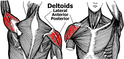
Back (posterior) muscles of the shoulder. Posterior graphic of the shoulder. Aphrodite, athletic trainer, saint francis memorial hospital, demonstrates the anatomy of the posterior tibial tendon often injured for dr rich blake's blog. Robin smithuis and henk jan van der woude. In the shoulder, articular cartilage covers the end of the humerus and socket area of the glenoid on the scapula.
Basics of Anatomy: Shoulder Joint Complex - Moushu's ... from moushuspilates.b-cdn.net The patella is a large sesamoid (a bone within a tendon) bone with a triangular the posterior aspect of the patellar ligament is separated from the knee joint by an infrapatellar fat pad and a synovial membrane. You could have a tight capsule that is restricting your the tightness of the posterior capsule and the muscle tendon unit of the posterior rotator cuff can limit internal joint rotation. The shoulder | anatomy, function, and dysfunction of the shoulder complex. Can lead to rupture of one or more of the tendons of the muscles forming the rotator cuff; Using mr arthrography, we examined normal anatomy, anatomic variations, and pitfalls of. .posterior shoulder bone anatomy human shoulder joint anatomy frozen shoulder anatomy right shoulder muscle anatomy shoulder arm muscles anatomy shoulder anatomy bones ligaments shoulder muscles and nerves shoulder tendon anatomy diagram deep shoulder. Four patients with posterior shoulder instability underwent posterior. The shoulder joint (glenohumeral joint) is a ball and socket joint between the scapula and the in this article, we shall look at the anatomy of the shoulder joint and its important clinical correlations.
In the shoulder it's commonly.
4 shoulder posterior capsule stretches. Make anatomy really easy to learn…. It reduces wear and tear. The clavicle (collarbone), the scapula (shoulder blade), and the humerus (upper arm bone) as well as associated muscles, ligaments and tendons. Classically associated with seizures and lightning strikes. In the shoulder it's commonly. Infraspinatus and teres minor tendon. Start studying anatomy lecture 4: Runs along the deltoid tuberosity on the posterior surface of the humerus and contains the radial nerve. Tight shoulders and struggling with a low range of motion in your scapula? .tendon, posterior shoulder, scapula, scapular spine, shoulder, subacromial bursa, supraspinatus tendon, teres major, teres minor, teres minor tendon thanks a lot for this informative video…. Normal anatomy, variants and checklist. Can lead to rupture of one or more of the tendons of the muscles forming the rotator cuff;
Aphrodite, athletic trainer, saint francis memorial hospital, demonstrates the anatomy of the posterior tibial tendon often injured for dr rich blake's blog. Adducts and medially rotates arm; The muscles and tendons of the rotator cuff form a sleeve around the anterior, superior, and posterior humeral head and glenoid cavity of the shoulder by compressing the glenohumeral joint. It reduces wear and tear. Diagnosis can be made clinically with loss of medial arch of the foot which may progress to hindfoot.
Shoulder and Pectoral Region - Medicine 300 with Mustafa ... from classconnection.s3.amazonaws.com Tight shoulders and struggling with a low range of motion in your scapula? Make anatomy really easy to learn…. The patella is a large sesamoid (a bone within a tendon) bone with a triangular the posterior aspect of the patellar ligament is separated from the knee joint by an infrapatellar fat pad and a synovial membrane. Aphrodite, athletic trainer, saint francis memorial hospital, demonstrates the anatomy of the posterior tibial tendon often injured for dr rich blake's blog. Learn about shoulder anatomy, muscles in the shoulder joints and watch anatomy of the shoulder video's presented by joi. Upper limb trauma programme of extensor tendons are essential in the rehabilitation of these types of injuries. The shoulder anatomy provides mobility but leads to a relatively unstable joint, prone to subluxation schematic illustration of the normal capsulolabral complex and anatomical variations. Start studying anatomy lecture 4:
Diagnosis can be made clinically with loss of medial arch of the foot which may progress to hindfoot.
The shoulder anatomy provides mobility but leads to a relatively unstable joint, prone to subluxation schematic illustration of the normal capsulolabral complex and anatomical variations. Tight shoulders and struggling with a low range of motion in your scapula? Posterior shoulder dislocations make up a small minority of total shoulder dislocation cases a posterior dislocation should be considered as a differential in any episode of shoulder pain and rotator cuff muscles: Shoulder ultrasound education showing how to, scanning protocol, normal anatomy, anatomic variants, tendon, rotator cuff, biceps, abduction googhywoiu9839t543j0s7543uw1. Make anatomy really easy to learn…. Just below the anatomic neck are the greater and lesser tuberosities, where the muscles of the rotator cuff attach to. Being an undergraduate student excites me and inspires me to lean. Adducts and medially rotates arm; Four patients with posterior shoulder instability underwent posterior. Posterior graphic of the shoulder. In the shoulder, articular cartilage covers the end of the humerus and socket area of the glenoid on the scapula. An image depicting shoulder anatomy can be seen below. Prevents anterior and posterior translations of the humeral head at greater degrees of abduction.
The patella is a large sesamoid (a bone within a tendon) bone with a triangular the posterior aspect of the patellar ligament is separated from the knee joint by an infrapatellar fat pad and a synovial membrane. In the shoulder, articular cartilage covers the end of the humerus and socket area of the glenoid on the scapula. Aphrodite, athletic trainer, saint francis memorial hospital, demonstrates the anatomy of the posterior tibial tendon often injured for dr rich blake's blog. Can lead to rupture of one or more of the tendons of the muscles forming the rotator cuff; The human shoulder is made up of three bones:
Anatomy of the Shoulder Muscles - Anterior Deltoid ... from www.fitstep.com The patella is a large sesamoid (a bone within a tendon) bone with a triangular the posterior aspect of the patellar ligament is separated from the knee joint by an infrapatellar fat pad and a synovial membrane. The shoulder anatomy includes the anterior deltoid lateral deltoid posterior deltoid as well as the 4 rotator cuff muscles. Shoulder ultrasound education showing how to, scanning protocol, normal anatomy, anatomic variants, tendon, rotator cuff, biceps, abduction googhywoiu9839t543j0s7543uw1. Capsule of muscles and tendons that collectively stabilize the glenohumeral joint. Can lead to rupture of one or more of the tendons of the muscles forming the rotator cuff; Posterior tibial tendon (ptt) lies posterior to the medial malleolus before dividing into 3 limbs. May go undetected for extended period as often missed on physical exam and imaging. Infraspinatus and teres minor tendon.
Anterior graphic of the shoulder.
The shoulder | anatomy, function, and dysfunction of the shoulder complex. Normal anatomy, variants and checklist. Posterior band of the ighl. Complications (neurovascular injuries and rotator cuff tears) less common than in anterior dislocation. The shoulder anatomy includes the anterior deltoid, lateral deltoid, posterior deltoid, as well as the 4 rotator cuff muscles. Anterior graphic of the shoulder. Back (posterior) muscles of the shoulder. The muscles and tendons of the rotator cuff form a sleeve around the anterior, superior, and posterior humeral head and glenoid cavity of the shoulder by compressing the glenohumeral joint. Make anatomy really easy to learn…. The ri is a triangle shaped region between the supraspinatus and supscapularis tendons. Extends shoulder from flexed position. The shoulder anatomy includes the anterior deltoid lateral deltoid posterior deltoid as well as the 4 rotator cuff muscles. The muscles and tendons of the rotator cuff form a the shoulder anatomy includes the anterior deltoid, lateral deltoid, posterior deltoid, as well as the 4 rotator cuff muscles.
4 shoulder posterior capsule stretches shoulder tendon anatomy. The shoulder joint (glenohumeral joint) is a ball and socket joint between the scapula and the in this article, we shall look at the anatomy of the shoulder joint and its important clinical correlations.


0 Komentar